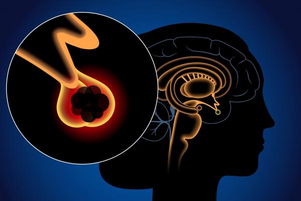
Surgical Treatments Applied in the Pituitary Diseases
Cases with a pituitary disease in the clinic are presented to the council, which consists of experienced specialists at Yeditepe University Koşuyolu Hospital. The patients, whose surgical treatment is decided by the decision of the council, are taken into detailed preoperative examination by the endocrinologist, neuroradiologist, neuro-ophthalmologist, neuro anesthesiologist and neurosurgeon.
Following the necessary preparations in terms of anesthesia, the patient is admitted to the clinic. The patient and his/her relatives are informed about the technique of the surgical treatment, the method of intervention, its risks, the postoperative process, and alternative treatment methods, and their consent is obtained.
Surgical Treatments Applied in the Pituitary Diseases Clinic
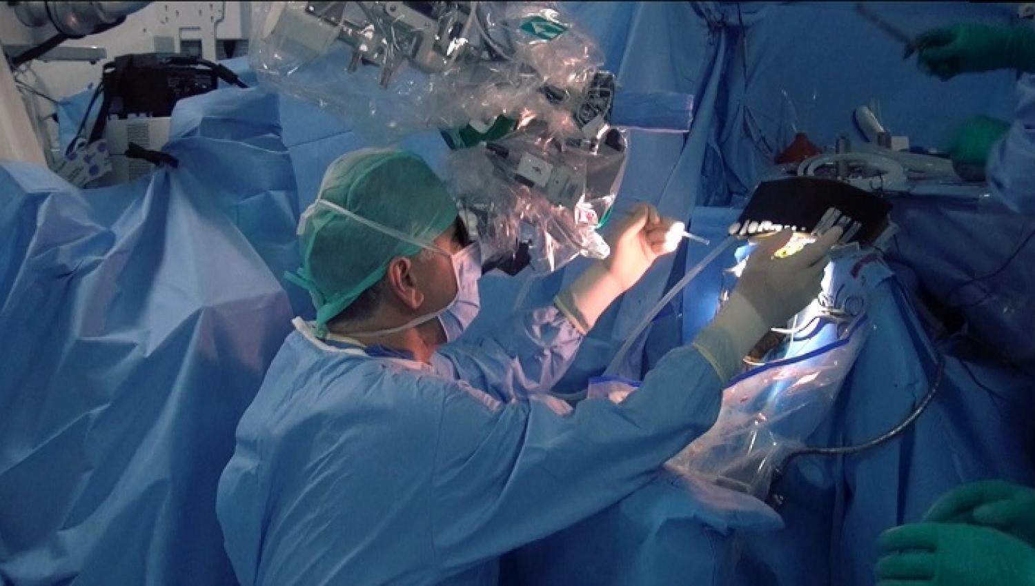
Endoscope-assisted Microneurosurgery and Transsphenoidal Approaches are used most frequently in the surgical treatment of pituitary tumors in our clinic. It is aimed to radically remove the tumors, and for this purpose, the following advanced technology devices are used in combination.
Surgical Microscope: By using appropriate microsurgical techniques in neurosurgery operations, it enables the operation to be performed in a smaller area with less touching of normal tissue. Thanks to the surgical microscope, the surgeon can see the operating area much better by using smaller instruments and therefore can work more safely. In addition to creating a safe treatment option for the patient, it provides the opportunity to perform operations with smaller incisions, thus shortening the time of the patient's discharge and return to his/her normal life.
In order to perform microsurgery, a microscope that provides focusing and magnification at different depths, microsurgical instruments suitable for working under the microscope, and most importantly, experience in this subject is required. In our hospital, tumors in the pituitary region can be treated by entering through the nose in appropriate cases with surgical microscopes equipped with up-to-date technology.
Neuroendoscope: It is an auxiliary tool frequently used in micro neurosurgery that contributes to the diagnosis and treatment of brain lesions under HD or 4K camera images. With its variable-angle optics system that can enter the surgical site and the high resolution it provides, regions that cannot be visualized with a surgical microscope can be visualized. The neuro endoscope, which is used successfully in appropriate cases, helps to understand the relationship of the lesion with the surrounding tissues during the operation, to confirm the removal of the entire lesion, and to provide bleeding control in the surgical site. For these reasons, the use of intraoperative neuro endoscopes in surgery for pituitary lesions provides significant gains in terms of surgical treatment success rate.
Intraoperative Ultrasound (ioUSG): It is a dynamic imaging method that provides simultaneous visual information with a high resolution during surgical procedures. With ioUSG, the location of the tumor and its relationship with the surrounding tissues are determined precisely. It guides the surgeon in important issues such as the determination of the entrance site to the tumor during the operation and the determination of whether the tumor has been completely removed. ioUSG can be used in both transcranial and transsphenoidal procedures. Intraoperative USG, which requires experience to use, is superior to other imaging systems because it is fast, provides simultaneous images, and does not affect the displacement of the brain. It is possible to obtain safer and more successful treatment results with the use of intraoperative USG in appropriate cases.
Neuronavigation: It is a set of computer-assisted technologies that show the localization and relationship with the surrounding tissues for the safe removal of brain and spinal cord tumors. Image-guided neuronavigation systems have been developed to assist in more reliable resection of brain tumors. In appropriate cases, using neuronavigation tools, the location of the lesion is determined more clearly before the surgery and the surgical strategy is determined accordingly.
Intraoperative MRI (ioMRI): It is a system that allows MR imaging of the surgical site and its surroundings at any stage of the surgery. In appropriate cases, it can be used to evaluate the integrity and dynamic changes in the lesion and surrounding tissues. ioMRI requires the use of specially designed operating rooms and MRI-compatible surgical devices. With this MR imaging performed during the surgery, the surgeon obtains the most accurate information about the lesion and distinguishes the abnormal brain tissue from the normal brain tissue. The quality of the MRI device used is important in making this distinction. Thanks to the 3-Tesla MRI used in our clinic, the tumor and surrounding tissues can be visualized in detail.
In Yeditepe University Pituitary Diseases Clinic, patients are evaluated by our specialist team and referred for the most appropriate treatment option.
All patients referred for surgical treatment are closely followed up by a multidisciplinary team before, during, and after surgery.
In Yeditepe University Pituitary Diseases Clinic, surgery for pituitary diseases that require close follow-up before, during, and after surgery is performed at international standards with an experienced team and up-to-date technological infrastructure.
”
See Also

Relevant Services