If You Have Diabetes, Consult An Ophthalmologist At Least Once A Year.
Prof. Dr. Sinan Tatlıpınar, Head of the Ophthalmology Department at Yeditepe University Hospital, explains...
What is Diabetic Retinopathy?
Diabetic Retinopathy is the most common of the eye diseases related to diabetes. It is a prominent cause of vision loss in adults. Uncontrolled diabetes affects the eye vessels as well as the whole body. It damages the vessels in the retinal layer responsible for vision. As a result, the blood in the vessel leaks out and causes edema in the retina. As the disease progresses, abnormal new vessels may form on the retinal surface and cause intraocular hemorrhages. The risk of developing the disease increases with the duration of diabetes. The disease usually affects both eyes.
How Many Stages Are There in Diabetic Retinopathy?
It has four stages.
Mild retinopathy: It is the earliest stage. Small bubbles (micro-aneurysm) begin to form on the walls of small blood vessels in the retina.
Moderate retinopathy: As the disease progresses, some vessels that feed the retina become occluded.
Severe retinopathy: With the increase in the number of occluded vessels in this stage, areas that cannot be fed appear in the retina. New vessel formation is stimulated by sending some signals from the areas with impaired feeding.
Proliferative retinopathy: It is the most advanced stage. As a result of the signals sent by the retina for vessel formation, new vessels appear. The new vessels are abnormal and bleed very easily. They sometimes grow onto the surface of the retina and sometimes into the transparent gel (vitreus) that fills the inside of the eye. When these veins bleed, severe vision loss occurs.
Does Diabetic Retinopathy Adversely Affect the Eyes of Every Diabetic?
There is a risk of diabetic retinopathy for all patients with type 1 and type 2 diabetes. For this reason, everyone with diabetes should have a comprehensive fundus examination with the dilated pupil at least once a year. The longer you have diabetes, the greater your risk of developing retinopathy.
What Can Be Done to Protect Vision?
As previously mentioned, if you have diabetes, have a detailed fundus examination at least once a year. If you have been diagnosed with retinopathy, you may need more frequent checkups. With early diagnosis, treatment, and regular follow-up, the risk of blindness can be significantly reduced. Control your blood sugar well. Thus, you can slow the emergence and progression of retinopathy.
Does Diabetic Retinopathy Cause Symptoms?
Usually, in the early stages of the disease, the patient does not have any complaints. Blurred vision may occur if fluid leaks into the macula (the area of the retina where sharp vision is provided). If abnormal new vessels form on the retinal surface and these vessels cause intraocular hemorrhage, sudden vision loss occurs.
Is Bleeding in Diabetic Retinopathy Noticeable From The Outside?
First, you may see spots of blood or floating spots in the eye. If you develop spots in your eyes, consult your ophthalmologist as soon as possible. You may need treatment before more serious bleeding occurs. Sometimes without treatment, the spots may disappear and you may see better. However, bleeding may recur and severe visual impairment may occur. You should be examined by your ophthalmologist before bleeding increases. Otherwise, severe vision loss may occur. The earlier the treatment is administered, the greater the chance of success.
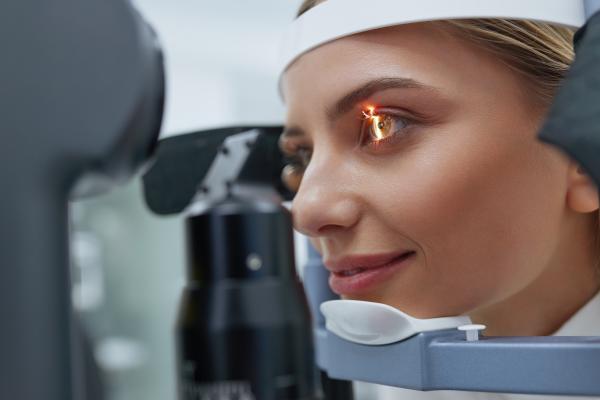
How Is Diabetic Retinopathy Diagnosed?
Visual acuity test: The level of vision is measured. Eye examination by dilating the pupil: Drops are put into your eye to dilate your pupil. In this way, your ophthalmologist can scan larger areas inside your eye and search for signs of the disease. Using a special lens, your doctor investigates whether there is any damage to the retina of your eye and your optic nerve.
Ocular angiography and retinal tomography (OCT): These are tests used to evaluate the bottom of the eye in more detail.
Is Diabetic Retinopathy Difficult to Treat?
In the first 3 stages of the disease, there is no need for treatment in the process that there is no macular edema. Stage 4 is treated with a laser. The laser aims to help close the abnormal blood vessels. It is usually completed in 2 or more sessions. The laser is more effective if applied before the abnormal blood vessels begin to bleed. If the bleeding is severe, an operation called a vitrectomy may be needed. With this surgery, the blood in the eye is cleaned.
How Is Macular Edema Treated?
One of the main treatments is laser. This method is called focal laser therapy. If there is edema in both eyes and laser treatment is required, it is usually applied to one eye first and then to the other eye. Focal laser therapy helps to preserve existing vision. In macular edema that does not respond to laser, special medications are given with a small needle into the eye. It is usually done in the operating room and takes a very short time. In some cases, resistant to this method, vitrectomy surgery can be used in the treatment of edema.
”
See Also
Related Doctors


Relevant Services